
X-ray, CT, MRI
17
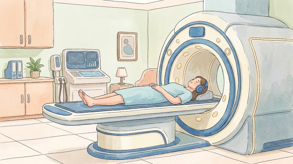
Chest X-rayA picture of your lungs and heart taken with X-rays. The radiation dose is small.⏱ usually 5 to 10 minutesChest X-ray screeningA quick image of your lungs for a routine checkup. The radiation dose is very small.⏱ usually 2 to 5 minutesBone and joint X-rayA picture of your bones and joints taken with X-rays. It shows fractures, cracks, and changes in the joints.⏱ usually 5 to 15 minutesSpine X-rayAn X-ray picture of your spine. It shows the bones and vertebrae. The radiation dose is small.⏱ usually 5 to 15 minutesPanoramic dental X-ray (OPG)One picture that shows all your teeth, jaws, and roots. The machine moves around your head. The radiation dose is small.⏱ usually 5 minutes, the picture itself about 15 secondsMammogramAn X-ray of the breasts. Each breast is pressed between plates for a few seconds so the picture is clear.⏱ usually 10 to 20 minutesBone density scan (DEXA)A special X-ray that measures how dense your bones are. The radiation dose is very small.⏱ usually 10 to 20 minutesHead CTA CT scan shows your brain and skull bones in thin slices. It is fast, and usually no needle is needed.⏱ usually 5 to 10 minutes in the room; the scan itself takes less than a minuteChest CTA CT scan shows your lungs in thin slices, in much more detail than a regular x-ray. Usually no needle is needed.⏱ usually 5 to 10 minutes in the room; the scan itself takes a few secondsCT scan of the abdomen with contrastA CT scan shows the organs in your belly in thin slices. Contrast dye given into a vein makes blood vessels and organs easier to see.⏱ 15 to 30 minutes in the scan room, sometimes longer if you need to drink contrast firstCT angiographyA CT scan of your blood vessels. Contrast dye is injected into a vein, and the machine takes pictures that show how blood flows through your vessels.⏱ usually 15 to 30 minutes with preparation; the scan itself takes a few minutesBrain MRIPictures of the brain taken in a magnetic field. There is no X-ray radiation. Sometimes contrast is given into a vein.⏱ usually 20 to 40 minutes, longer with contrastSpine MRIPictures of the spine, discs, and nerves taken in a magnetic field. There is no radiation. Usually one part is scanned: the neck, the upper back, or the lower back.⏱ usually 20 to 40 minutes for one part of the spineJoint MRIPictures of a joint taken in a magnetic field. They show ligaments, menisci, cartilage, and muscles. There is no radiation.⏱ usually 20 to 30 minutesAbdominal and pelvic MRIA machine with a strong magnet takes detailed pictures of the organs in your belly and pelvis. There is no X-ray radiation.⏱ usually 30 to 60 minutes, longer with contrastBarium X-ray of the stomach and bowelYou drink a white liquid with barium, and the X-ray shows your food pipe, stomach, and bowel. Barium is not absorbed and leaves with your stool.⏱ usually 20 to 40 minutes; if the small bowel is checked, images are taken over several hoursPET-CT scanA small amount of radioactive sugar is given into a vein. The scanner shows where cells take it up most actively and combines this with a CT image.⏱ usually 2 to 3 hours at the center; the scan itself takes 15 to 30 minutes