
← All services · X-ray, CT, MRI
Bone and joint X-ray
joint X-ray · arm X-ray · leg X-ray · spine X-ray · bone X-ray
A picture of your bones and joints taken with X-rays. It shows fractures, cracks, and changes in the joints.
After an injury, for joint or back pain, or to check how a fracture is healing.
⏱ Duration: usually 5 to 15 minutes
Be sure to tell your doctor
How to prepare
- Preparation. None needed for arms, legs, and joints. You can eat and drink.
- Lower back and tailbone. Sometimes you are asked to eat fewer gas-causing foods the day before and to empty your bowels. Check with your clinic.
- Metal. Take off jewelry, watches, and anything metal from the part of the body being X-rayed.
What to bring
- Your referral
- Past X-rays of the same body part, to compare
How it goes
- You uncover the body part being checked
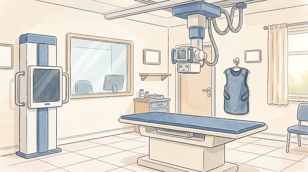
- The radiographer places it on the table or plate in the right position
- The rest of your body may be covered with a protective apron
- You need to keep still for a couple of seconds while the picture is taken
- Usually 2 pictures are taken in different positions
What you'll feel
- You do not feel the X-ray itself
- After an injury, holding the position can hurt. Tell the radiographer
Afterwards
- You can go back to your day straight away
- The images and report are usually ready the same day
- Take the results to the doctor who referred you
Contact a doctor straight away if
- After an injury, the arm or leg turns blue, goes numb, swells a lot, or the bone looks out of shape: call emergency services
Common questions
Can an X-ray show ligaments and the meniscus?
No. An X-ray shows bones. Ligaments, menisci, and cartilage are seen better on MRI or ultrasound.
Why are two pictures taken?
A fracture can hide on one picture. Two positions give an accurate view.