
← All services · X-ray, CT, MRI
Joint X-ray
joint X-ray · knee X-ray · shoulder X-ray · X-ray of a joint
An X-ray of a joint. It shows the bones, the joint space, fractures, and signs of wear.
After an injury, for pain, swelling, clicking, or limited movement in a joint, or to check after treatment.
⏱ Duration: usually 5 to 15 minutes in the room; the report the same or the next day
Be sure to tell your doctor
How to prepare
- Prep. None needed. You can eat and drink.
- Clothing. Take off jewelry and anything metal in the area. Sometimes you are asked to change.
What to bring
- Your referral
- Past X-rays of this joint, for comparison
How it goes
- You uncover the joint and remove jewelry
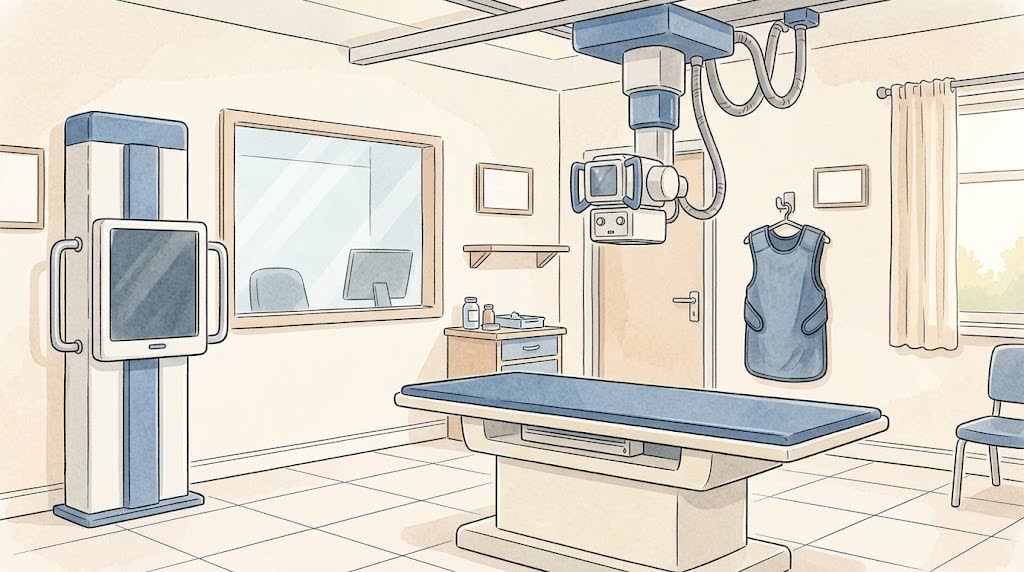
- The technician places the joint in the right position
- Usually two images are taken from different sides
- Sometimes both joints are imaged, or while standing under your weight
- Each time, you keep still for a couple of seconds
What you'll feel
- It does not hurt. The image takes a fraction of a second
- Sometimes you need to hold an awkward position for a bit
- With a fresh injury, positioning can hurt. Tell the technician
Afterwards
- You can go about your day right away
- Take the images and report to an orthopedic or trauma doctor
Common questions
Does an X-ray show ligaments and the meniscus?
No, X-rays show bones. Ligaments and menisci are seen better on MRI or ultrasound.
Why image the healthy joint?
Sometimes to compare it with the painful one. Your doctor decides.