
Joint ultrasound
knee ultrasound · shoulder ultrasound · joint ultrasound
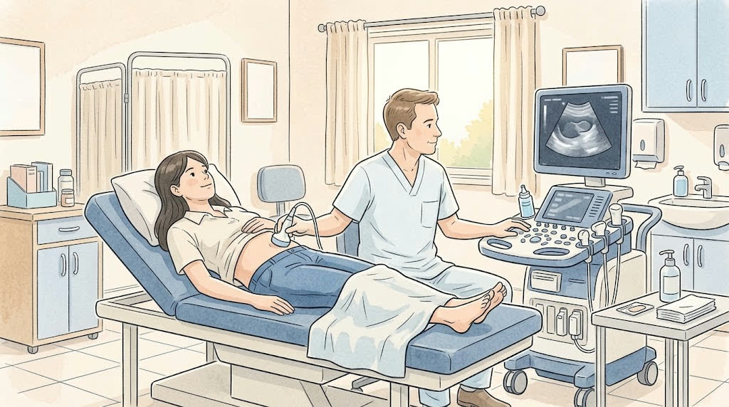
The doctor uses ultrasound to look at the joint, ligaments, tendons, and muscles. It is safe and uses no radiation.
For joint pain, swelling, or injury, or if a torn ligament or fluid in the joint is suspected.
⏱ Duration: usually 15 to 30 minutes
Be sure to tell your doctor
How to prepare
- Preparation. No special preparation is needed. You can eat and drink as usual.
- Clothing. Wear something comfortable so the joint is easy to uncover. For a knee scan, wear shorts or loose pants.
- Skin. Do not put cream or ointment on that area on the day of the scan.
What to bring
- Your referral
- Past images and reports: X-ray, MRI, ultrasound
How it goes
- You uncover the joint and sit or lie down
- The doctor puts on cool gel
- The probe is moved over the skin around the joint
- You will be asked to bend, straighten, or turn your arm or leg
- Often the other joint is checked too, for comparison
What you'll feel
- The gel is cool
- If the joint is sore, pressure from the probe may be unpleasant. Tell the doctor
Afterwards
- The gel is wiped off, and you can go back to your day right away
- You usually get the report right away
Common questions
Which is better for a joint, ultrasound or MRI?
Ultrasound shows ligaments, tendons, and fluid well, and it can be done while you move. MRI shows deeper parts better, like the meniscus and cartilage. The doctor decides.
Can ultrasound show bones?
Only their surface. For bones and fractures, you need an X-ray or CT scan.